Thursday, March 3, 2011

Mobile Pet Vacc Schedule

Endocrine System Nervous System Central Nervous System


4. nerves


6. brain


9. The cerebral peduncles and colliculi


12. striatum


1. Introduction


The Nervous System, the most complete and unknown to all who make up the human body says with the Endocrine System, the functions of the organismo.Capaz control to receive and integrate numerous data from different sensory organs to achieve a response from the body, the nervous system usually is responsible for monitoring the activities faster. In addition, the nervous system is responsible for the intellective functions, such as memory the emotions or volitions.


Your anatomical constitution is very complex, and cells that compose it, unlike the rest of the body, lack regenerative capacity.


Next will have to know everything related to the Central Nervous System .


2. Fundamentals of the nervous system and its functions


The human being is endowed with neural mechanisms through which it receives information of the alterations that occur in your external and internal environment and others, that able to react to the information properly. Through these mechanisms see and hear, act, analyze, organize and store in your brain records of their experiences.


These neural mechanisms lines are set communication system calls nerviosoEl whole nervous system is divided into: central nervous system


includes:


Brain.


Spinal Cord.
Also called "Life on" because their functions are:


perceive stimuli from the outside world.


transmit nerve impulses to sensory processing centers.


production or effector impulses government.


effector transmission of these impulses to skeletal muscles.


peripheral nervous system


includes:


Cranial


Nerve function raquídeos.Tiene as receiving and transmitting to the system central nervous sensory impulses, and to target organs other impulses .


autonomic nervous system


includes:


sympathetic trunk, formed by nerve cords that extend longitudinally along the neck, thorax and abdomen on each side of spine.


peripheral lymph . (The nodes are groups of cell bodies).

This system is called also "autonomous." Is related to the viscera, glands, heart , blood vessels and lisos.Su muscles is efferent function, transmitting impulses that regulate the functions of the organs in accordance with the vital requirements of the moment.


3. The neuron


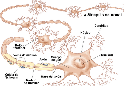
The neuron cell is nervous neuroblasto.Es derived from the functional unit of the nervous system provides a link for communicating between receptors and effectors, through nerve fibers.


consists of three parts:


body or soma, composed mainly of nucleus, cytoplasm and nucleolus.


Dendrites: nerve endings.


Axon: long ending, which can reach up to one meter in length.The axon usually has multiple endings called terminal buttons, which are in proximity to the dendrites or in the body of another neuron .


The separation between the axon of one neuron and the dendrites or the body of another, is of the order of 0.02 micras.Esta relationship between the axon of one neuron and the dendrites of another is called "synapses ".


Through synapses, a neuron sends a message pulses from axon to dendrites or other body, and transmitting information nerviosa.La synaptic transmission has the following character istics: The nerve impulse conduction takes place in one sense: the axon of a neuron body or dendrites of another neuron synapse. The nerve impulse is propagated through chemical intermediates, such as acetylcholine and norepinephrine, which are released by the axon terminals of the first neuron and are received by the following prompt in it the production of a new momentum. In the central nervous system neurons, excitatory and inhibitory and each substance releases its own mediator. The speed impulse conduction along nerve fibers varies from 1 to 100 meters per second, according to their size, being higher in the longest. When the presynaptic terminals are stimulated continuously or in high frequency, transmitted pulses decreased in number because of a "synaptic fatigue." The transmission of a signal from one neuron to another suffers a delay of 5 milliseconds.
The neuroglia. The central nervous system man is about 10 billion neurons and 5 to 10 times more glial cells.


These cells form a tissue called glia that has the following duties: Provide support to the brain and spinal cord. Skirting the blood vessels forming an impenetrable barrier to toxins. Neurons provide vital chemicals. Removed by phagocytosis, the dead tissue. Insulate axons through the myelin.


4. The nerves are nerviosLos usually do or sets of axons, but the sensory nerves which are composed of functional dendrites long ranging from the "boom" of the spinal cord to the sensory receptors and their function of conducting impulses as axons. The various nerve fibers forming a unity maintained by connective tissue. The nerves can be classified in several ways: By origin: spinal: Composed of nerve fibers of the anterior roots and motor and sensory or posterior roots, which leave the bone through the foramina. The spinal nerves are visceral and somáticosLos visceral elements are related to structures surrounding the digestive, respiratory, urogenital and vascular system and most of the glands.


somata are related to the coating body tissues and voluntary muscles.


Skull: There are 12 pairs of nerves that arise from the brainstem at the level of the fourth ventricle, above the medulla and serve mostly to specialized senses of the face and head. Its performance is mixed, ie, it contains sensory and motor fibers.


Among the cranial nerves are: the olfactory, the optic, which joins the central nervous system to the thalamus, the oculomotor, the trochlear or pathetic, the abducens, the trigeminal nerve fibers sensory temperature, pain, touch and pressure , the facial, the ramjet-acoustic; and acoustic receivers position and movements of the head, the glossopharyngeal, vagus and the spinal accessory halibut.


By function:


Sensory or afferent

:


conduct impulses that inform the various sensations.


motors or efferent:


conduct impulses to motor functions.


Mixed: fibers contain sensory and motor fibers.


For collectors: exteroceptive: For pulses produced by outside stimuli body: touch, temperature , pain, pressure , and sensory organs like the eye and ear .


proprioceptive

: For stimuli born in the same body: muscles, tendons, joints and related balance. Interoceptive: For the impulses from the viscera: digestive system, respiratory, circulatory, urogenital and glands.


5. The spinal cord


The spinal cord is a cylindrical mass of nerve tissue that occupies the spinal canal is 40 to 45 cm in length and extends from the foramen magnum, which is continuous with the medulla to the lumbar region.
is protected by the meningeal membranes, pia, arachnoid and dura mater and cerebrospinal fluid.
From the region of the second lumbar vertebra, where the bone ends, to rump, down a filament delgadollamado "filum terminale" and sacral nerve roots and lumbar vertebrae, forming a bundle of fibers called "tail horse. "
From out 31 pairs of spinal nerves that give it a segmented: 8 cervical, 12 thoracic, 5 lumbar, 5 sacral, and coccygeal.
Bone consists of a gray matter consists of cell bodies, and white matter consists of myelinated fibers ascending and descending.
ascending fibers are ascending beams that are sensitive and conduct impulses they receive from skin, muscles and joints to different brain areas.
descending fibers are descending beams are engines and conduct impulses from higher centers of the brain others who reside in the bone or the muscles and glands.
The gray matter has a widening called "horns", two gift dorsal or later, two ventral and two intermediate or above and are located between the dorsal and ventral. The dorsal horns contain neurons that control motor responses of the autonomic nervous system and the ventral motor neurons whose axons end in the muscles of the somatic system.
In the center of the gray matter and along it there is a small fluid-filled canal cefalorraquídeo.Otro important aspect of the spinal anatomy, is that there are neurons which connect between the sensory and motor fibers, which gives rise reflex responses that need not be ordered by the brain centers.
The functions of a bone are
center is a partnership, through which are made reflexes.
is a two-way direction :
from the periphery to the brain centers (sensitive).
From brain centers to the periphery (motor).
6. The brain
The brain is the central nervous system enclosed in the cranial cavity.
is divided into:
Forebrain.
midbrain. Brain
later.


The brain or rhombencephalon posterior is located in the part immediately above the spinal cord and consists of three structures : the medulla, pons or bridge, and the cerebellum. It is also the fourth ventricle.


The forebrain or prosencephalon is divided into diencephalon and telencephalon.
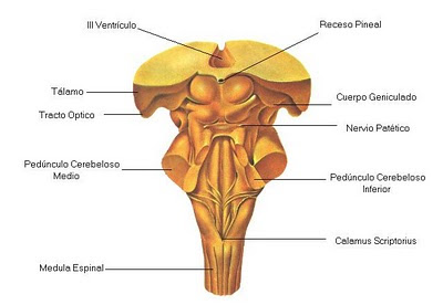
The diencephalon comprises: the thalamus, hypothalamus, optic chiasm, the pituitary, the mamillary bodies and the cavity called the third ventricle.
the telencephalon consists of the basal ganglia: caudate and lenticular nuclei that form the corpus striatum and the amygdaloid body and faculty, the rhinencephalon, hippocampus and septal area, which form the limbic system, and the cerebral cortex or neocortex . The enlargement of the telencephalon
form the cerebral hemispheres consisting of three lobes: frontal, temporal and occipital lobes. Externamente los hemisferios tienen múltiples pliegues separados por hendiduras que cuando son profundas se llaman cisuras.
Los dos hemisferios están unidos por el cuerpo calloso, formado por fibras que cruzan de un hemisferio a otro.
La corteza cerebral es una capa de sustancia gris que se extiende sobre la superficie de los hemisferios.
De estas estructuras del encéfalo sólo vamos a estudiar algunas que tienen importancia más resaltante para comprender las bases fisiológicas de la conducta .
7. El bulbo
Es una estructura que se halla en el extremo superior de la médula y como prolongación de ella. En el hombre mide unos 3 cm de longitud. A bulb
level crossing some nerve bundles going to the opposite side of the brain after join those who had crossed in the medulla. Similarly fibers coming from the brain across the bulb to go to the opposite side through the bone.
Bulb Features:
is the most important center of vegetative life because in it are located the central connections related breathing and heart rate, any injury can be fatal in this region.
connection serves some cranial nerves.
The bulb is involved in the following reflections: VO myth, coughing, salivation, the breathing, sneezing, sucking, swallowing, and vasomotor.

Iit Chemistry /o.p.agarwal


The nervous system is a network of highly specialized tissues, whose main component to neurons, cells that are interconnected in complex ways that have the ability to drive, using electrochemical signals (see Synapse), a variety of stimuli within the nervous tissue and to most other tissues, and coordinating multiple functions in the body. DivisionesAnatómicamente, the nervous system Homo sapiens (as well as many other species) are grouped in different organs, which actually make up stations where they spend the neural pathways. Thus, for study, these bodies can be grouped according to their location, in two parts: central nervous system and peripheral nervous system.
The Central Nervous System : This comprises the Brain and Spinal Cord , is protected by three membranes, the meninges . Inside there is a system of cavities called ventricles, through which circulates the liquid cerebrospinal . The brain is the central nervous system is protected by the skull . It comprises the brain the cerebellum and brainstem . Brain
: is the most voluminous. Is divided into two hemispheres, one right and one left, separated by the interhemispheric fissure and communicated through the corpus callosum . The surface is called cerebral cortex and consists of refolding called convolutions formed gray. Underlying it is the white matter. In deep zones are areas of gray matter forming nuclei and the thalamus the caudate nucleus or hypothalamus .
Cerebellum: It's in the bottom and back of the brain, lodged in the posterior cranial fossa near the brainstem.
Brainstem : Composed of midbrain the pons and medulla . Connects the brain to the spinal cord .
The spinal cord is an extension the brain, like a cord that extends inside the spine . It is the gray matter inside and white outside.
Peripheral Nervous System: It consists of the nerves, cranial and spinal nerves that emerge from the central nervous system and travel throughout the body, containing axons of neural pathways with different functions and the lymph peripherals that are in the path containing nerves and cell bodies, the only outside the central nervous system. The
cranial nerves are 12 pairs that send sensory information from the neck and head to the central nervous system. Receive motor commands to control the skeletal muscles of the neck and head. The
spinal nerves are 31 pairs and are in charge of sending sensory information (touch, pain and temperature) of the trunk and limbs and the position and condition of the muscles and joints of the trunk and limbs to the central nervous system and, from there, receiving motor commands for the control of skeletal muscle which are driven by the spinal division espinal.Una less anatomical but much more functional, which divides the system nervous according to the role that different neural pathways, regardless of whether they walk part of the central nervous system or peripheral.
The somatic nervous system : Also called the nervous system of personal relationships, is formed by the set of neurons that regulate voluntary or conscious functions in the body (eg muscle movement, touch).
The autonomic nervous system, also called the autonomic nervous system or (incorrectly) visceral nervous system is formed by the set of neurons that regulate involuntary or unconscious functions in the body (eg bowel movement, visceral sensitivity).
noteworthy that neurons in both systems can reach or leave these bodies if they are voluntary and involuntary functions (and, indeed, these organs are the majority.) In some texts it is considered that the autonomic nervous system is part of the peripheral nervous system, but this is incorrect because, in its way, some neurons in the autonomic nervous system can be passed by both the central nervous system by the peripheral, which also occurs in the somatic nervous system. The division between central and peripheral nervous system has only anatomical purposes.
In turn, the vegetative system is classified into sympathetic and parasympathetic , systems whose functions are largely antagonistic. We have in our body approximately 150,000 miles of nerves that run throughout our body.

Pay For Your Own Dinner Invitation

Excretion

excretion in humans
-excretion is the last process of the role of nutrition, but just as important as everyone else. The unit responsible for carrying out the excretory, but also involved other devices diferentes.El excretory apparatus is responsible for cleaning the blood of waste products it has collected in each tissue and organ. It consists of the kidneys, ureters, bladder and urethra.
The kidneys are the organs responsible for cleansing the blood of waste, making the urine as a final product. have about 12 cm in length and are arranged in the back of abdomen.Los ureters are the tubes that start from the renal pelvis and carries urine to the bladder.
The bladder is a muscular organ, shaped like a balloon, which expands as it fills with urine and compressed in the act of urination. The bladder capacity is approximately 350 cm3. When the tension of the walls of the bladder exceeds a certain value, there is a nervous reflex, which receives Reflecting the name of urination, and urination becomes conscious.
The urethra is a tube from the bladder and the urine is expelled by urination.
The kidneys are made up of nephrons, which are responsible for producing urine. In a nephron can distinguish the following parts: glomerulus, proximal tubule, loop of Henle, distal tubule and collecting duct, which collects the urine.

Crm Basic Exam Anwsers

Watson and Crick article


MOLECULAR STRUCTURE OF NUCLEIC ACIDS
A Structure for Deoxyribonucleic Acid
We suggest a structure for the salt of deoxyribonucleic acid (DNA .). This structure has novel features which are of considerable biological interest.
A structure for nucleic acid has been proposed by Pauling and Corey (1). Have kindly put at our disposal the manuscript before publication. Their model consists of three intertwined chains, with phosphates near the axis of the fiber, and the bases out. In our opinion, this structure is unsatisfactory for two reasons: (1) believe that the material obtained the X-ray diagrams is the salt, not the free acid. Without the acidic hydrogen atoms is not clear what forces can hold the structure together, especially as the negatively charged phosphates near the axis will repel each other. (2) Some of the van der Waals distances appear to be too small.
Another triple chain structure has been suggested by Fraser (in press). In his model the phosphates are out and the bases inward, held together by hydrogen bonds. This structure thus described is rather ill-defined as not discussed.
We offer here a radically different structure for the salt deoxyribonucleic acid. This structure has two helical chains each lap around the same axis (see diagram). We made the usual chemical assumptions, specifically, that each chain consists of phosphate diester groups joining beta-D residues desoxirribofuranosa links-3 ', 5'. The two chains (but not their bases) are related by a dyad perpendicular to the axis of the fiber. Both chains follow right-handed helix, but due to the dyad the sequences of atoms in the two chains run in opposite directions. Each of the chains separate model resembles Furberg No. 1 (2), that is, the bases are on the inside of the loop and phosphates on the outside. The configuration of the sugar and the atoms near approaches to the "standard configuration" of Furberg, sugar is available at right angles to the base attached. There is a residue on each chain every 3.4 Å in the direction-z. We have assumed an angle of 36 degrees between adjacent residues in the same chain, so that the structure is repeated after 10 residues on each chain, ie after 34 Å. The distance of a phosphorus atom from the axis of the fiber is 10 Å. As the phosphates are on the outside, cations have easy access to them. The structure is open and the water content is rather high. For us, content low bases and the structure would approach would be more compact.
The novel aspect of the structure is the manner in which the two chains are held together by purine and pyrimidine bases. The planes of the bases are perpendicular to the axis of the fiber. They meet in pairs, a base of one of the chains connected by hydrogen bonds to a base of the other chain, and thus the two are joined side by side with identical z-coordinate One of the pair must be purine and one pyrimidine. Hydrogen bonds are as follows: purine position 1 to pyrimidine position 1; purine position 6 to pyrimidine position 6 [etc.]
This figure is purely schematic. The two ribbons symbolize the sugar-phosphate chains, and the horizontal rods the pairs of bases holding the chains together. The vertical line marks the axis of the fiber.

Assuming that the bases only occur within the structure in the most plausible tautomeric forms (that is, setting the keto rather than enol) are the specific pairs of bases can join. These pairs are: adenine (purine) with thymine (pyrimidine) and guanine (purine) with cytosine (pyrimidine).
In other words, if an adenine is a member of a couple, on a string, then the other member must be thymine; something similar happens for the guanine and cytosine. The sequence of bases on a single chain does not appear to be restricted in any way. However, if they can only form specific base pairs, it follows that knowing the sequence of bases on one strand, then the sequence on the other chain is automatically determined.
has been found experimentally (3.4) that the ratio of adenine to thymine, and the ratio of guanine to cytosine, are always very close to unity for deoxyribonucleic acid. It is probably impossible to build this structure with a ribose sugar instead of deoxyribose, the extra oxygen atom would make too closed and form a van der Waals bond.
The X-ray data previously published (5.6) on deoxyribonucleic acid are insufficient for a rigorous test of our structure. So far we can say is roughly compatible with the experimental data, but should be seen as unproven until you have verified with more accurate results. Some of these will be submitted on the following communications. We were not aware of the details of the results presented when we devised our structure, which rests mainly but not entirely on published and experimental data and stereochemical arguments.
not escaped our communication pairing específico que hemos postulado sugiere inmediatamente un mecanismo copiador para el material genético .
Todos los detalles de la estructura, incluyendo las condiciones presumidas para su construcción, junto con un conjunto de coordenadas para los átomos, se publicarán con posterioridad.
Estamos en deuda con el Dr. Jerry Donohue por las constantes críticas y consejos, especialmente sobre distancias interatómicas. También hemos sido estimulados por el conocimiento general de la naturaleza y los resultados experimentales inéditos así como ideas del Dr. M.H.F. Wilkins , la Dra. R.E. Franklin y sus colaboradores del King's College , en Londres. Uno Whom (JDW) has been funded by a grant from the National Foundation for infantile paralysis.
JD Watson FHC Crick

Medical Research Council Unit for the Study of the Structure Molecular Biological Systems
Cavendish Laboratory, Cambridge


April 2 (1) Pauling, L. and Corey, RB, Nature, 171, 346 (1953); Proc.USNat.Sci., 39, 84 (1953).
(2) Furberg, S. Minutes Chem.Scand., 6, 634 (1952).
(3) Chargaff, e. For reference see Zamenhof, S, Brawerman, G. and Chargaff, E., Biochem. et. Biophys. Ata, 9, 402 (1952).
(4) Wyatt, GR, J. Gen.Physiol., 36, 201 (1952).
(5) Astbury, WT, Symp.Soc.Exp.Biol. 1, Nucleic Acid, 66 (Cambridge University Press, 1947).
(6) Wilkins, MHF and Randall, JT, Biochim. et Biophys. Acta, 10, 192 (1953).

Article published in the journal Nature , April 25, 1953, p. 737.